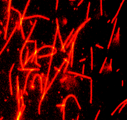
Tubulin (X-rhodamine labeled; Bovine Brain micr-injctn) 微管蛋白(X-罗丹明标记;牛脑micr注射)
X-rhodamine microtubules formed from X-rhodamine labeled tubulin.
Product Uses Include
Laser based applications
Monitoring microtubule dynamcs in living cells
Speckle microscopy
Formation of fluorescent microtubules
Microscopy studies of MAP and microtubule associated motor activities
Nanotechnology
Material
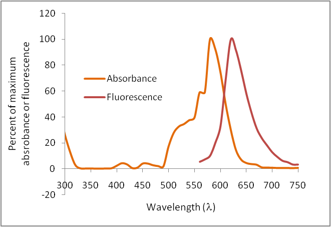
Bovine brain tubulin (>99% pure, see Cat. # T240) has been modified to contain covalently linked X-rhodamine at random surface lysines. An activated ester of X-rhodamine was used to label the protein. Labeling stoichiometry was determined by spectroscopic measurement of protein and dye concentrations (dye extinction coefficient when protein bound is 66,000M-1cm-1). Final labeling stoichiometry is 1-2 dyes per tubulin heterodimer. X-rhodamine labeled tubulin can be detected using a filter set of 540-560 nm excitation and 610-630 emission. X-rhodamine tubulin is in a versatile, stable and easily shipped format. It is ready for micro-injection or in vitro polymerization. Cytoskeleton, Inc. also offers AMCA (Cat. # TL440M), HiLyte Fluor™ 488TM (Cat. # TL488M), rhodamine (Cat. # TL590M) and HiLyte Fluor™ 647TM (Cat. # TL670M) labeled tubulins of the same quality.
Purity
The protein purity of the tubulin used for labeling is determined by scanning densitometry of Coomassie Blue stained protein on a 4-20% polyacrylamide gel. The protein used for TL620M is >99% pure tubulin (Figure 1 A). Labeled protein is run on an SDS gel and photographed under green light. Any unincorporated X-rhodamine dye would be visible in the dye front. No fluorescence is detected in the dye front, indicating that no free dye is present in the final product (Figure 1 B).